by George Taniwaki
After my consultation with the surgeon, I go to the radiology lab to have a computed tomographic scan (CT scan or CAT scan) of my abdomen taken. This scan will produce a 3D image of my kidneys and the arteries and veins that connect to it.
The surgeon and nephrologist will review this image to ensure that I have two functioning kidneys and select which one I can safely donate.
If both kidneys are the same size, they will want to remove the left one, since it is easier to get to. If they are not the same size, they will want to remove the smaller one (and leave me with the bigger one).
Finally, if the arteries and veins don’t branch a lot, they can use a technique called laparoscopic surgery. Unlike traditional open surgery, where the incision must be large enough for the surgeon to insert his/her hand into the body, laparoscopic surgery uses one or more small incisions and the operation is performed using thin robot instruments inserted through the holes, called ports. This reduces the chance of bleeding and infection. When successful, it also leads to shorter recovery time, less scarring, and less postoperative pain.
| What is it? | CT scan angiogramCreates a 3D image of the arteries and other structures in the abdomen |
| Why is it needed? | Ensures you have two functioning kidneys and that one can be safely removed. Also helps the surgeon plan the operation |
| How is it done? | A contrast agent is added to the blood and a series of x-rays are taken |
| Preparation | None |
| Test time | One hour |
| Risks | Repeated exposure to x-ray energy can increase the chance of cancer. A CT scan is the equivalent of about 40 chest x-rays |
| Discomfort | You have to pull down your pants (you can leave your underwear on). The contrast agent can leave a metallic taste in your mouth and can cause tingling in your abdomen and bladder. The test is not painful |
How a CT scan image is created
A CT scan is a series of thin sliced cross-section images of an object, in this case my abdomen. The images are created by sending a large number of x-ray beams through the object from various X-Y directions and measuring the energy absorbed from each beam. By using an algorithm, called the inverse Radon transform, the data from all the beams are combined to produce the density value (shades from black to white) for each pixel, or more accurately each voxel, and produces a single X-Y slice. By moving the object (my body) back and forth along the Z-axis, a large number of slices can be created and the entire 3D object can be imaged.
The procedure
The CT scanner at UWMC is a GE model. It consists of a large ring, about five feet in diameter. The x-ray source and the detector are attached to a rotating ring while the patient lies on a non-metallic table that can slide back and forth through the center of the ring.
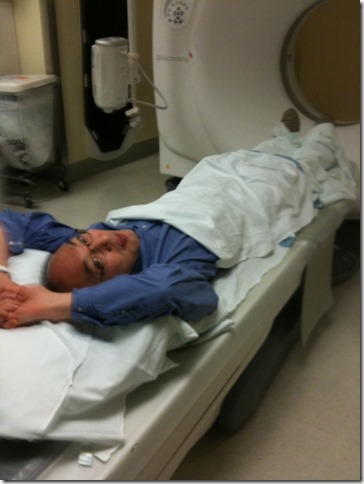
Conveniently, I don’t have to disrobe and put on a silly hospital gown for this procedure. I just lay on the sliding table. The operator covers me with a sheet and I slide my pants down to my knees (so that the metal zipper doesn’t interfere with the scan). I have to raise my hands above my head so that they don’t interfere with the scan either.
Ready for a CT scan. Photo by UWMC staff
The scan consists of three passes. In the first pass, a low-resolution image is taken to ensure that my abdomen is in the correct position. After this initial pass, the operator returns from behind the lead wall and attaches a small IV drip of iodine solution to my arm for about 1 minute. The iodine will absorb x-ray energy, which will increase the contrast of the image. It will make blood vessels like the renal arteries stand out to produce an image called an angiogram. Iodine is also filtered out of the blood by the kidneys, and the amount of iodine collected by each kidney will indicate how well each kidney is functioning.
Even though the contrast agent is injected into my arm, it causes my mouth to feel dry and my abdomen to feel warm and tingly. The operator tells me that some patients say the feeling is similar to urinating on oneself. Two high-resolution passes are made after the iodine IV. One immediately after the IV that will show the iodine in my arteries and another about 6 minutes later after the kidneys have filtered the iodine from my blood and it collects in my bladder. (I had no idea that the kidneys worked so fast.)
The images
After completing the CT scan, I walk past a sign that says the fee for copying radiological images onto CDs and shipping them to another hospital is $13.50. Being curious, I ask if I can just pay the fee and take the CDs with me. The clerk says sure and so I ask how I can view the images. He says the viewer app is on the CDs. Then he asks me if I know how to interpret the images. I say no, but that’s OK, I’ll figure it out on my own.
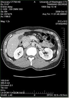
When I get home, I load the three CDs containing almost 1,500 images of slices of my body, start the viewer app, and start clicking on the controls. A few samples are shown below. Very cool! I only wish the images were in pseudocolor (like red for iodine, gray for everything else) and in solid 3D so you could spin them around.
CT scans of my abdomen. Images from UWMC
Top left: Front or coronal view of lower abdomen before the iodine injection (low resolution pass), this view is synthesized by combining multiple cross-sectional slices (notice that my left kidney, on the right of the image, is higher and larger than the right one, also notice the slight curvature of my spine that may indicate I have a very mild case of scoliosis)
Top right: Cross-section of my upper abdomen immediately after iodine injection (notice how the iodine absorbs the x-rays making the heart show as white while the lungs, which contain little blood, show as black)
Bottom left: Cross-section of my lower abdomen immediately after iodine injection (my kidneys are the two white blobs, again notice that my left kidney, which is on the right of the image, is larger than the right one)
Bottom right: Same cross-section 6 minutes later (notice how the bladder located between the kidneys turns white as it fills with iodine, the kidneys are even whiter than before, but the other organs fade to gray as the iodine is filtered from the blood)
For more information on becoming a kidney donor, see my Kidney donor guide.
[Update: It is the contrast agent, not the x-rays that caused the tingling sensation. Also, the scanner makes 3 passes, not 4.]


November 15, 2009 at 2:45 pm
You know what else would be cool? Being able to view these images on an iPhone. Maybe I’ll write an app for that.